Back Chromosoom Afrikaans Chromosom ALS ሃብለበራሂ Amharic صبغي Arabic كروموزوم ARY Cromosoma AST Xromosom Azerbaijani کوروموزوم AZB Хромосома Bashkir Храмасома Byelorussian
| Part of a series on |
| Genetics |
|---|
 |

- Chromatid
- Centromere
- Short arm
- Long arm
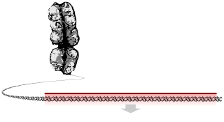
A chromosome is a package of DNA containing part or all of the genetic material of an organism. In most chromosomes, the very long thin DNA fibers are coated with nucleosome-forming packaging proteins; in eukaryotic cells, the most important of these proteins are the histones. Aided by chaperone proteins, the histones bind to and condense the DNA molecule to maintain its integrity.[1][2] These eukaryotic chromosomes display a complex three-dimensional structure that has a significant role in transcriptional regulation.[3]
Normally, chromosomes are visible under a light microscope only during the metaphase of cell division, where all chromosomes are aligned in the center of the cell in their condensed form.[4] Before this stage occurs, each chromosome is duplicated (S phase), and the two copies are joined by a centromere—resulting in either an X-shaped structure if the centromere is located equatorially, or a two-armed structure if the centromere is located distally; the joined copies are called 'sister chromatids'. During metaphase, the duplicated structure (called a 'metaphase chromosome') is highly condensed and thus easiest to distinguish and study.[5] In animal cells, chromosomes reach their highest compaction level in anaphase during chromosome segregation.[6]
Chromosomal recombination during meiosis and subsequent sexual reproduction plays a crucial role in genetic diversity. If these structures are manipulated incorrectly, through processes known as chromosomal instability and translocation, the cell may undergo mitotic catastrophe. This will usually cause the cell to initiate apoptosis, leading to its own death, but the process is occasionally hampered by cell mutations that result in the progression of cancer.
The term 'chromosome' is sometimes used in a wider sense to refer to the individualized portions of chromatin in cells, which may or may not be visible under light microscopy. In a narrower sense, 'chromosome' can be used to refer to the individualized portions of chromatin during cell division, which are visible under light microscopy due to high condensation.
- ^ Hammond CM, Strømme CB, Huang H, Patel DJ, Groth A (March 2017). "Histone chaperone networks shaping chromatin function". Nature Reviews. Molecular Cell Biology. 18 (3): 141–158. doi:10.1038/nrm.2016.159. PMC 5319910. PMID 28053344.
- ^ Wilson, John (2002). Molecular biology of the cell : a problems approach. New York: Garland Science. ISBN 978-0-8153-3577-1.
- ^ Bonev, Boyan; Cavalli, Giacomo (14 October 2016). "Organization and function of the 3D genome". Nature Reviews Genetics. 17 (11): 661–678. doi:10.1038/nrg.2016.112. hdl:2027.42/151884. PMID 27739532. S2CID 31259189.
- ^ Alberts B, Bray D, Hopkin K, Johnson A, Lewis J, Raff M, Roberts K, Walter P (2014). Essential Cell Biology (Fourth ed.). New York, New York, US: Garland Science. pp. 621–626. ISBN 978-0-8153-4454-4.
- ^ Schleyden, M. J. (1847). Microscopical researches into the accordance in the structure and growth of animals and plants. Printed for the Sydenham Society.
- ^ Antonin W, Neumann H (June 2016). "Chromosome condensation and decondensation during mitosis" (PDF). Current Opinion in Cell Biology. 40: 15–22. doi:10.1016/j.ceb.2016.01.013. PMID 26895139.
