Back قشرة حزامية Arabic Xiru cinguláu AST Cingulární kortex Czech Gyrus cinguli German Giro cingulado Spanish Vöökäär Estonian قشر سینگولیت Persian Gyrus cingulaire French פיתול החגורה HE 帯状回 Japanese
| Cingulate cortex | |
|---|---|
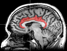
 Medial surface of left cerebral hemisphere, with cingulate gyrus and cingulate sulcus highlighted. | |
| Details | |
| Part of | Cerebral cortex |
| Artery | Anterior cerebral |
| Vein | Superior sagittal sinus |
| Identifiers | |
| Latin | cortex cingularis, gyrus cinguli |
| Acronym(s) | Cg |
| MeSH | D006179 |
| NeuroNames | 159 |
| NeuroLex ID | birnlex_798 |
| TA98 | A14.1.09.231 |
| TA2 | 5513 |
| FMA | 62434 |
| Anatomical terms of neuroanatomy | |


The cingulate cortex is a part of the brain situated in the medial aspect of the cerebral cortex. The cingulate cortex includes the entire cingulate gyrus, which lies immediately above the corpus callosum, and the continuation of this in the cingulate sulcus. The cingulate cortex is usually considered part of the limbic lobe.
It receives inputs from the thalamus and the neocortex, and projects to the entorhinal cortex via the cingulum. It is an integral part of the limbic system, which is involved with emotion formation and processing,[1] learning,[2] and memory.[3][4] The combination of these three functions makes the cingulate gyrus highly influential in linking motivational outcomes to behavior (e.g. a certain action induced a positive emotional response, which results in learning).[5] This role makes the cingulate cortex highly important in disorders such as depression[6][7] and schizophrenia.[8] It also plays a role in executive function and respiratory control.

- ^ Hadland, K. A.; Rushworth M.F.; et al. (2003). "The effect of cingulate lesions on social behaviour and emotion". Neuropsychologia. 41 (8): 919–931. doi:10.1016/s0028-3932(02)00325-1. PMID 12667528. S2CID 16475051.
- ^ "Cingulate binds learning". Trends Cogn Sci. 1 (1): 2. 1997. doi:10.1016/s1364-6613(97)85002-4. PMID 21223838. S2CID 33697898.
- ^ Kozlovskiy, S.; Vartanov A.; Pyasik M.; Nikonova E.; Velichkovsky B. (10 October 2013). "Anatomical Characteristics of Cingulate Cortex and Neuropsychological Memory Tests Performance". Procedia - Social and Behavioral Sciences. 86: 128–133. doi:10.1016/j.sbspro.2013.08.537.
- ^ Kozlovskiy, S.A.; Vartanov A.V.; Nikonova E.Y.; Pyasik M.M.; Velichkovsky B.M. (2012). "The Cingulate Cortex and Human Memory Processes". Psychology in Russia: State of the Art. 5: 231–243. doi:10.11621/pir.2012.0014.
- ^ Hayden, B. Y.; Platt, M. L. (2010). "Neurons in Anterior Cingulate Cortex Multiplex Information about Reward and Action". Journal of Neuroscience. 30 (9): 3339–3346. doi:10.1523/JNEUROSCI.4874-09.2010. PMC 2847481. PMID 20203193.
- ^ Drevets, W. C.; Savitz, J.; Trimble, M. (2008). "The subgenual anterior cingulate cortex in mood disorders". CNS Spectrums. 13 (8): 663–681. doi:10.1017/s1092852900013754. PMC 2729429. PMID 18704022.
- ^ Cite error: The named reference
:0was invoked but never defined (see the help page). - ^ Adams, R.; David, A. S. (2007). "Patterns of anterior cingulate activation in schizophrenia: A selective review". Neuropsychiatric Disease and Treatment. 3 (1): 87–101. doi:10.2147/nedt.2007.3.1.87. PMC 2654525. PMID 19300540.