Back طور النمو الثاني Arabic G2 faza BS G2 fáze Czech Fase G2 Spanish فاز G2 Persian G2 (podfaza) Croatian Fase G2 Italian G2期 Japanese G2-фаза Kazakh Faza G2 Polish


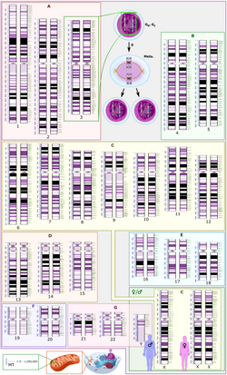
G2 phase, Gap 2 phase, or Growth 2 phase, is the third subphase of interphase in the cell cycle directly preceding mitosis. It follows the successful completion of S phase, during which the cell’s DNA is replicated. G2 phase ends with the onset of prophase, the first phase of mitosis in which the cell’s chromatin condenses into chromosomes.
G2 phase is a period of rapid cell growth and protein synthesis during which the cell prepares itself for mitosis. Curiously, G2 phase is not a necessary part of the cell cycle, as some cell types (particularly young Xenopus embryos[1] and some cancers[2]) proceed directly from DNA replication to mitosis. Though much is known about the genetic network which regulates G2 phase and subsequent entry into mitosis, there is still much to be discovered concerning its significance and regulation, particularly in regards to cancer. One hypothesis is that the growth in G2 phase is regulated as a method of cell size control. Fission yeast (Schizosaccharomyces pombe) has been previously shown to employ such a mechanism, via Cdr2-mediated spatial regulation of Wee1 activity.[3] Though Wee1 is a fairly conserved negative regulator of mitotic entry, no general mechanism of cell size control in G2 has yet been elucidated.
Biochemically, the end of G2 phase occurs when a threshold level of active cyclin B1/CDK1 complex, also known as Maturation promoting factor (MPF) has been reached.[4] The activity of this complex is tightly regulated during G2. In particular, the G2 checkpoint arrests cells in G2 in response to DNA damage through inhibitory regulation of CDK1.
- ^ Alberts B, Johnson A, Lewis J, Raff M, Roberts K, Walter P (2002). "An Overview of the Cell Cycle". Molecular Biology of the Cell (4th ed.). New York: Garland Science. ISBN 978-0-8153-3218-3.
- ^ Liskay RM (April 1977). "Absence of a measurable G2 phase in two Chinese hamster cell lines". Proceedings of the National Academy of Sciences of the United States of America. 74 (4): 1622–5. Bibcode:1977PNAS...74.1622L. doi:10.1073/pnas.74.4.1622. PMC 430843. PMID 266201.)
- ^ Moseley JB, Mayeux A, Paoletti A, Nurse P (June 2009). "A spatial gradient coordinates cell size and mitotic entry in fission yeast". Nature. 459 (7248): 857–60. Bibcode:2009Natur.459..857M. doi:10.1038/nature08074. PMID 19474789. S2CID 4330336.
- ^ Sha W, Moore J, Chen K, Lassaletta AD, Yi CS, Tyson JJ, Sible JC (February 2003). "Hysteresis drives cell-cycle transitions in Xenopus laevis egg extracts". Proceedings of the National Academy of Sciences of the United States of America. 100 (3): 975–80. Bibcode:2003PNAS..100..975S. doi:10.1073/pnas.0235349100. PMC 298711. PMID 12509509.