Back خلية بينية Arabic লাইডিগ কোষ Bengali/Bangla Leydigove ćelije BS Cèl·lules de Leydig Catalan Leydigova buňka Czech Leydig-Zwischenzelle German Κύτταρα Λέυντιγκ Greek Célula de Leydig Spanish Leydigin solu Finnish Cellule de Leydig French
| Leydig cell | |
|---|---|
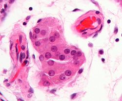
 Micrograph showing a cluster of Leydig cells (center of image). H&E stain. | |
 Histological section through testicular parenchyma of a boar. 1 Lumen of convoluted part of the seminiferous tubules, 2 spermatids, 3 spermatocytes, 4 spermatogonia, 5 Sertoli cell, 6 myofibroblasts, 7 Leydig cells, 8 capillaries | |
| Identifiers | |
| MeSH | D007985 |
| FMA | 72297 |
| Anatomical terms of microanatomy | |
Leydig cells, also known as interstitial cells of the testes and interstitial cells of Leydig, are found adjacent to the seminiferous tubules in the testicle and produce testosterone in the presence of luteinizing hormone (LH).[1][2] They are polyhedral in shape and have a large, prominent nucleus, an eosinophilic cytoplasm, and numerous lipid-filled vesicles.[3]
While Leydig cells are predominantly associated with testosterone production in the male testes, recent studies have revealed their presence and role in the ovaries of females. These cells, located in the ovarian interstitial tissue, contribute to the production of androgens, including testosterone, which plays a significant role in the regulation of the menstrual cycle, ovulation, and overall reproductive health in women. The activity of Leydig cells is particularly important in conditions such as polycystic ovary syndrome (PCOS), where excess androgen production can lead to symptoms such as hirsutism, acne, and infertility.
- ^ Chabner, Davi-Ellen (2016). The Language of Medicine - E-Book. Elsevier Health Sciences. p. 316. ISBN 978-0-32-337083-7.
- ^ Johnson, Martin H. (2018). Essential Reproduction. John Wiley & Sons. p. 131. ISBN 978-1-11-924645-9.
- ^ Zhou, Ming; Netto, George; Epstein, Jonathan I (2022). Uropathology E-Book. Elsevier Health Sciences. p. 428. ISBN 978-0-32-365396-1.