Back تجدد Arabic Regenerasiya Azerbaijani Регенерация Bulgarian Regeneracija (biologija) BS Regeneració Catalan Regenerace Czech Regeneration (Physiologie) German Αναγέννηση (βιολογία) Greek Regeneración (biología) Spanish Regeneratsioon Estonian


Regeneration in biology is the process of renewal, restoration, and tissue growth that makes genomes, cells, organisms, and ecosystems resilient to natural fluctuations or events that cause disturbance or damage.[1] Every species is capable of regeneration, from bacteria to humans.[2][3][4] Regeneration can either be complete[5] where the new tissue is the same as the lost tissue,[5] or incomplete[6] after which the necrotic tissue becomes fibrotic.[6]
At its most elementary level, regeneration is mediated by the molecular processes of gene regulation and involves the cellular processes of cell proliferation, morphogenesis and cell differentiation.[7][8] Regeneration in biology, however, mainly refers to the morphogenic processes that characterize the phenotypic plasticity of traits allowing multi-cellular organisms to repair and maintain the integrity of their physiological and morphological states. Above the genetic level, regeneration is fundamentally regulated by asexual cellular processes.[9] Regeneration is different from reproduction. For example, hydra perform regeneration but reproduce by the method of budding.
The regenerative process occurs in two multi-step phases: the preparation phase and the redevelopment phase.[10][11] Regeneration begins with an amputation which triggers the first phase. Right after the amputation, migrating epidermal cells form a wound epithelium which thickens, through cell division, throughout the first phase to form a cap around the site of the wound.[10] The cells underneath this cap then begin to rapidly divide and form a cone shaped end to the amputation known as a blastema. Included in the blastema are skin, muscle, and cartilage cells that de-differentiate and become similar to stem cells in that they can become multiple types of cells. Cells differentiate to the same purpose they originally filled meaning skin cells again become skin cells and muscle cells become muscles. These de-differentiated cells divide until enough cells are available at which point they differentiate again and the shape of the blastema begins to flatten out. It is at this point that the second phase begins, the redevelopment of the limb. In this stage, genes signal to the cells to differentiate themselves and the various parts of the limb are developed. The end result is a limb that looks and operates identically to the one that was lost, usually without any visual indication that the limb is newly generated.
The hydra and the planarian flatworm have long served as model organisms for their highly adaptive regenerative capabilities.[12] Once wounded, their cells become activated and restore the organs back to their pre-existing state.[13] The Caudata ("urodeles"; salamanders and newts), an order of tailed amphibians, is possibly the most adept vertebrate group at regeneration, given their capability of regenerating limbs, tails, jaws, eyes and a variety of internal structures.[2] The regeneration of organs is a common and widespread adaptive capability among metazoan creatures.[12] In a related context, some animals are able to reproduce asexually through fragmentation, budding, or fission.[9] A planarian parent, for example, will constrict, split in the middle, and each half generates a new end to form two clones of the original.[14]
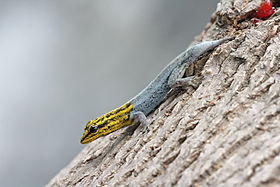
Echinoderms (such as the sea star), crayfish, many reptiles, and amphibians exhibit remarkable examples of tissue regeneration. The case of autotomy, for example, serves as a defensive function as the animal detaches a limb or tail to avoid capture. After the limb or tail has been autotomized, cells move into action and the tissues will regenerate.[15][16][17] In some cases a shed limb can itself regenerate a new individual.[18] Limited regeneration of limbs occurs in most fishes and salamanders, and tail regeneration takes place in larval frogs and toads (but not adults). The whole limb of a salamander or a triton will grow repeatedly after amputation. In reptiles, chelonians, crocodilians and snakes are unable to regenerate lost parts, but many (not all) kinds of lizards, geckos and iguanas possess regeneration capacity in a high degree. Usually, it involves dropping a section of their tail and regenerating it as part of a defense mechanism. While escaping a predator, if the predator catches the tail, it will disconnect.[19]
- ^ Birbrair A, Zhang T, Wang ZM, Messi ML, Enikolopov GN, Mintz A, Delbono O (August 2013). "Role of pericytes in skeletal muscle regeneration and fat accumulation". Stem Cells and Development. 22 (16): 2298–314. doi:10.1089/scd.2012.0647. PMC 3730538. PMID 23517218.
- ^ a b Carlson BM (2007). Principles of Regenerative Biology. Elsevier Inc. p. 400. ISBN 978-0-12-369439-3.
- ^ Gabor MH, Hotchkiss RD (March 1979). "Parameters governing bacterial regeneration and genetic recombination after fusion of Bacillus subtilis protoplasts". Journal of Bacteriology. 137 (3): 1346–53. doi:10.1128/JB.137.3.1346-1353.1979. PMC 218319. PMID 108246.
- ^ Sinigaglia, Chiara; Alié, Alexandre; Tiozzo, Stefano (2022), Blanchoud, Simon; Galliot, Brigitte (eds.), "The Hazards of Regeneration: From Morgan's Legacy to Evo-Devo", Whole-Body Regeneration, vol. 2450, New York, NY: Springer US, pp. 3–25, doi:10.1007/978-1-0716-2172-1_1, ISBN 978-1-0716-2171-4, PMC 9761548, PMID 35359300
- ^ a b Min S, Wang SW, Orr W (2006). "Graphic general pathology: 2.2 complete regeneration". Pathology. pathol.med.stu.edu.cn. Archived from the original on 2012-12-07. Retrieved 2012-12-07.
(1) Complete regeneration: The new tissue is the same as the tissue that was lost. After the repair process has been completed, the structure and function of the injured tissue are completely normal
- ^ a b Min S, Wang SW, Orr W (2006). "Graphic general pathology: 2.3 Incomplete regeneration". Pathology. pathol.med.stu.edu.cn. Archived from the original on 2013-11-10. Retrieved 2012-12-07.
The new tissue is not the same as the tissue that was lost. After the repair process has been completed, there is a loss in the structure or function of the injured tissue. In this type of repair, it is common that granulation tissue (stromal connective tissue) proliferates to fill the defect created by the necrotic cells. The necrotic cells are then replaced by scar tissue.
- ^ Himeno Y, Engelman RW, Good RA (June 1992). "Influence of calorie restriction on oncogene expression and DNA synthesis during liver regeneration". Proceedings of the National Academy of Sciences of the United States of America. 89 (12): 5497–501. Bibcode:1992PNAS...89.5497H. doi:10.1073/pnas.89.12.5497. PMC 49319. PMID 1608960.
- ^ Bryant PJ, Fraser SE (May 1988). "Wound healing, cell communication, and DNA synthesis during imaginal disc regeneration in Drosophila". Developmental Biology. 127 (1): 197–208. doi:10.1016/0012-1606(88)90201-1. PMID 2452103.
- ^ a b Brockes JP, Kumar A (2008). "Comparative aspects of animal regeneration". Annual Review of Cell and Developmental Biology. 24: 525–49. doi:10.1146/annurev.cellbio.24.110707.175336. PMID 18598212.
- ^ a b Kohlsdorf, Tiana; Schneider, Igor (March 2021). "Towards an evolutionary framework for animal regeneration". Journal of Experimental Zoology Part B: Molecular and Developmental Evolution. 336 (2): 87–88. Bibcode:2021JEZB..336...87K. doi:10.1002/jez.b.23034. ISSN 1552-5007. PMID 33600616. S2CID 231963500.
- ^ Tiozzo, Stefano; Copley, Richard R. (2015-06-23). "Reconsidering regeneration in metazoans: an evo-devo approach". Frontiers in Ecology and Evolution. 3. doi:10.3389/fevo.2015.00067. ISSN 2296-701X.
- ^ a b Sánchez Alvarado A (June 2000). "Regeneration in the metazoans: why does it happen?" (PDF). BioEssays. 22 (6): 578–90. doi:10.1002/(SICI)1521-1878(200006)22:6<578::AID-BIES11>3.0.CO;2-#. PMID 10842312. Archived from the original (PDF) on 2013-11-11. Retrieved 2010-12-15.
- ^ Reddien PW, Sánchez Alvarado A (2004). "Fundamentals of planarian regeneration". Annual Review of Cell and Developmental Biology. 20: 725–57. doi:10.1146/annurev.cellbio.20.010403.095114. PMID 15473858. S2CID 1320382.
- ^ Campbell NA (1996). Biology (4th ed.). California: The Benjamin Cummings Publishing Company, Inc. p. 1206. ISBN 978-0-8053-1940-8.
- ^ Wilkie IC (December 2001). "Autotomy as a prelude to regeneration in echinoderms". Microscopy Research and Technique. 55 (6): 369–96. doi:10.1002/jemt.1185. PMID 11782069. S2CID 20291486.
- ^ Maiorana VC (1977). "Tail autotomy, functional conflicts and their resolution by a salamander". Nature. 2265 (5594): 533–535. Bibcode:1977Natur.265..533M. doi:10.1038/265533a0. S2CID 4219251.
- ^ Maginnis TL (2006). "The costs of autotomy and regeneration in animals: a review and framework for future research". Behavioral Ecology. 7 (5): 857–872. doi:10.1093/beheco/arl010.
- ^ Edmondson, C. H. (1935). "Autotomy and regeneration of Hawaiian starfishes" (PDF). Bishop Museum Occasional Papers. 11 (8): 3–20.
- ^ "UCSB Science Line". scienceline.ucsb.edu. Retrieved 2015-11-02.