Back نفق فقري Arabic Canal vertebral Catalan دڕکە کەلێن CKB Páteřní kanál Czech Wirbelkanal German Canal espinal Spanish Lülisambakanal Estonian Orno-hodi Basque مجرای نخاعی Persian Canal vertébral French
| Spinal canal | |
|---|---|
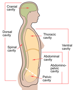
 Spinal cavity shown as part of dorsal body cavity. | |
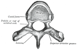
 A typical thoracic vertebra viewed from above. (Spinal canal is not labeled, but the foramen in the center would make up part of it.) | |
| Details | |
| Identifiers | |
| Latin | c. vertebralis |
| MeSH | D013115 |
| TA98 | A02.2.00.009 |
| TA2 | 1009 |
| FMA | 9680 |
| Anatomical terminology | |
In human anatomy, the spinal canal, vertebral canal or spinal cavity is an elongated body cavity enclosed within the dorsal bony arches of the vertebral column, which contains the spinal cord, spinal roots and dorsal root ganglia. It is a process of the dorsal body cavity formed by alignment of the vertebral foramina. Under the vertebral arches, the spinal canal is also covered anteriorly by the posterior longitudinal ligament and posteriorly by the ligamentum flavum. The potential space between these ligaments and the dura mater covering the spinal cord is known as the epidural space. Spinal nerves exit the spinal canal via the intervertebral foramina under the corresponding vertebral pedicles.
In humans, the spinal cord gets outgrown by the vertebral column during development into adulthood, and the lower section of the spinal canal is occupied by the filum terminale and a bundle of spinal nerves known as the cauda equina instead of the actual spinal cord, which finishes at the L1/L2 level.