Back غلالة باطنة Arabic Лумен (кръвоносен съд) Bulgarian Tunica intima German Túnica íntima Spanish درونلایه Persian Tunique intime d'un vaisseau sanguin French הגלימה הפנימית HE Tunika intima ID Tonaca intima Italian Интима Kazakh
This article includes a list of references, related reading, or external links, but its sources remain unclear because it lacks inline citations. (May 2015) |
| Tunica intima | |
|---|---|
 | |
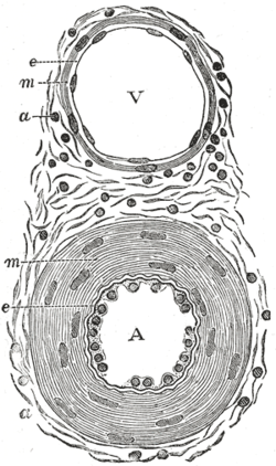
 Transverse section through a small artery and vein of the mucous membrane of the epiglottis of a child. (Tunica intima is at "e".) | |
| Details | |
| Part of | Wall of blood vessels |
| Identifiers | |
| Latin | tunica intima |
| MeSH | D017539 |
| TA98 | A12.0.00.018 |
| TA2 | 3922 |
| TH | H3.09.02.0.01003 |
| FMA | 55589 |
| Anatomical terminology | |
The tunica intima (Neo-Latin "inner coat"), or intima for short, is the innermost tunica (layer) of an artery or vein. It is made up of one layer of endothelial cells (and macrophages in areas of disturbed blood flow),[1][2] and is supported by an internal elastic lamina. The endothelial cells are in direct contact with the blood flow.
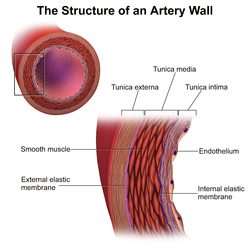
The three layers of a blood vessel are an inner layer (the tunica intima), a middle layer (the tunica media), and an outer layer (the tunica externa).
In dissection, the inner coat (tunica intima) can be separated from the middle (tunica media) by a little maceration, or it may be stripped off in small pieces; but, because of its friability, it cannot be separated as a complete membrane. It is a fine, transparent, colorless structure which is highly elastic, and, after death, is commonly corrugated into longitudinal wrinkles.
- ^ Scipione, Corey A.; Hyduk, Sharon J.; Polenz, Chanele K.; Cybulsky, Myron I. (December 2023). "Unveiling the Hidden Landscape of Arterial Diseases at Single-Cell Resolution". Canadian Journal of Cardiology. 39 (12): 1781–1794. doi:10.1016/j.cjca.2023.09.009. PMID 37716639.
- ^ Scipione, Corey A.; Cybulsky, Myron I. (October 2022). "Early atherogenesis: new insights from new approaches". Current Opinion in Lipidology. 33 (5): 271–276. doi:10.1097/MOL.0000000000000843. ISSN 0957-9672. PMC 9594136. PMID 35979994.