Back Aar Afrikaans Vena AN وريد Arabic ܘܪܝܕܐ ARC সিৰা Assamese Vena AST Бидурихь AV Vena Azerbaijani قارا دامار AZB Вена (анатомия) Bashkir
This article about biology may be excessively human-centric. (November 2024) |
| Vein | |
|---|---|
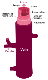
 Structure of a vein, which consists of three main layers: an outer layer of connective tissue, a middle layer of smooth muscle, and an inner layer lined with endothelium. | |
| Details | |
| System | Circulatory system |
| Identifiers | |
| Latin | vena |
| MeSH | D014680 |
| TA98 | A12.0.00.030 A12.3.00.001 |
| TA2 | 3904 |
| FMA | 50723 |
| Anatomical terminology | |
Veins (/veɪn/) are blood vessels in the circulatory system of humans and most other animals that carry blood towards the heart. Most veins carry deoxygenated blood from the tissues back to the heart; exceptions are those of the pulmonary and fetal circulations which carry oxygenated blood to the heart. In the systemic circulation, arteries carry oxygenated blood away from the heart, and veins return deoxygenated blood to the heart, in the deep veins.[1]
There are three sizes of veins: large, medium, and small. Smaller veins are called venules, and the smallest the post-capillary venules are microscopic that make up the veins of the microcirculation.[2] Veins are often closer to the skin than arteries.
Veins have less smooth muscle and connective tissue and wider internal diameters than arteries. Because of their thinner walls and wider lumens they are able to expand and hold more blood. This greater capacity gives them the term of capacitance vessels. At any time, nearly 70% of the total volume of blood in the human body is in the veins.[3] In medium and large sized veins the flow of blood is maintained by one-way (unidirectional) venous valves to prevent backflow.[3][1] In the lower limbs this is also aided by muscle pumps, also known as venous pumps that exert pressure on intramuscular veins when they contract and drive blood back to the heart.[4]
- ^ a b Moore HM, Gohel M, Davies AH (October 2011). "Number and location of venous valves within the popliteal and femoral veins: a review of the literature". J Anat. 219 (4): 439–43. doi:10.1111/j.1469-7580.2011.01409.x. PMC 3196749. PMID 21740424.
- ^ Guven G, Hilty MP, Ince C (2020). "Microcirculation: Physiology, Pathophysiology, and Clinical Application". Blood Purif. 49 (1–2): 143–150. doi:10.1159/000503775. PMC 7114900. PMID 31851980.
- ^ a b "Classification & Structure of Blood Vessels | SEER Training". training.seer.cancer.gov. Retrieved 29 January 2023.
- ^ GRAYS 2016, p. 131.