Back Seldeling Afrikaans انقسام الخلية Arabic Hüceyrənin bölünməsi Azerbaijani Күҙәнәк бүленеүе Bashkir কোষ বিভাজন Bengali/Bangla Ćelijska dioba BS Divisió cel·lular Catalan Buněčné dělení Czech Пайрăм пайланăвĕ CV Cellraniad Welsh


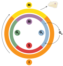
Cell division is the process by which a parent cell divides into two daughter cells.[1] Cell division usually occurs as part of a larger cell cycle in which the cell grows and replicates its chromosome(s) before dividing. In eukaryotes, there are two distinct types of cell division: a vegetative division (mitosis), producing daughter cells genetically identical to the parent cell, and a cell division that produces haploid gametes for sexual reproduction (meiosis), reducing the number of chromosomes from two of each type in the diploid parent cell to one of each type in the daughter cells.[2] Mitosis is a part of the cell cycle, in which, replicated chromosomes are separated into two new nuclei. Cell division gives rise to genetically identical cells in which the total number of chromosomes is maintained. In general, mitosis (division of the nucleus) is preceded by the S stage of interphase (during which the DNA replication occurs) and is followed by telophase and cytokinesis; which divides the cytoplasm, organelles, and cell membrane of one cell into two new cells containing roughly equal shares of these cellular components. The different stages of mitosis all together define the M phase of an animal cell cycle—the division of the mother cell into two genetically identical daughter cells.[3] To ensure proper progression through the cell cycle, DNA damage is detected and repaired at various checkpoints throughout the cycle. These checkpoints can halt progression through the cell cycle by inhibiting certain cyclin-CDK complexes. Meiosis undergoes two divisions resulting in four haploid daughter cells. Homologous chromosomes are separated in the first division of meiosis, such that each daughter cell has one copy of each chromosome. These chromosomes have already been replicated and have two sister chromatids which are then separated during the second division of meiosis.[4] Both of these cell division cycles are used in the process of sexual reproduction at some point in their life cycle. Both are believed to be present in the last eukaryotic common ancestor.
Prokaryotes (bacteria and archaea) usually undergo a vegetative cell division known as binary fission, where their genetic material is segregated equally into two daughter cells, but there are alternative manners of division, such as budding, that have been observed. All cell divisions, regardless of organism, are preceded by a single round of DNA replication.
For simple unicellular microorganisms such as the amoeba, one cell division is equivalent to reproduction – an entire new organism is created. On a larger scale, mitotic cell division can create progeny from multicellular organisms, such as plants that grow from cuttings. Mitotic cell division enables sexually reproducing organisms to develop from the one-celled zygote, which itself is produced by fusion of two gametes, each having been produced by meiotic cell division.[5][6] After growth from the zygote to the adult, cell division by mitosis allows for continual construction and repair of the organism.[7] The human body experiences about 10 quadrillion cell divisions in a lifetime.[8]
The primary concern of cell division is the maintenance of the original cell's genome. Before division can occur, the genomic information that is stored in chromosomes must be replicated, and the duplicated genome must be cleanly divided between progeny cells.[9] A great deal of cellular infrastructure is involved in ensuring consistency of genomic information among generations.[10][11][12]
- ^ Martin EA, Hine R (2020). A dictionary of biology (6th ed.). Oxford: Oxford University Press. ISBN 9780199204625. OCLC 176818780.
- ^ Griffiths AJ (2012). Introduction to genetic analysis (10th ed.). New York: W.H. Freeman and Co. ISBN 9781429229432. OCLC 698085201.
- ^ "10.2 The Cell Cycle – Biology 2e | OpenStax". openstax.org. 28 March 2018. Retrieved 2020-11-24.
- ^ Gilbert, Scott F. (2000), "Meiosis", Developmental Biology. 6th edition, Sinauer Associates, retrieved 2023-09-08
- ^ Gilbert SF (2000). "Spermatogenesis". Developmental Biology (6th ed.). Sinauer Associates.
- ^ Gilbert SF (2000). "Oogenesis". Developmental Biology (6th ed.). Sinauer Associates.
- ^ Maton, Anthea (1997). Cells : building blocks of life (3rd ed.). Upper Saddle River, N.J.: Prentice-Hall. pp. 70–74. ISBN 978-0134234762. OCLC 37049921.
- ^ Quammen D (April 2008). "Contagious Cancer". Harper's Magazine. ISSN 0017-789X. Retrieved 2019-04-14.
- ^ Golitsin, Yuri N.; Krylov, Mikhail C. C. (2010). Cell division : theory, variants, and degradation. New York: Nova Science Publishers. p. 137. ISBN 9781611225938. OCLC 669515286.
- ^ Fletcher, Daniel A.; Mullins, R. Dyche (28 January 2010). "Cell mechanics and the cytoskeleton". Nature. 463 (7280): 485–492. Bibcode:2010Natur.463..485F. doi:10.1038/nature08908. ISSN 0028-0836. PMC 2851742. PMID 20110992.
- ^ Li, Shanwei; Sun, Tiantian; Ren, Haiyun (27 April 2015). "The functions of the cytoskeleton and associated proteins during mitosis and cytokinesis in plant cells". Frontiers in Plant Science. 6: 282. doi:10.3389/fpls.2015.00282. ISSN 1664-462X. PMC 4410512. PMID 25964792.
- ^ Hohmann, Tim; Dehghani, Faramarz (18 April 2019). "The Cytoskeleton—A Complex Interacting Meshwork". Cells. 8 (4): 362. doi:10.3390/cells8040362. ISSN 2073-4409. PMC 6523135. PMID 31003495.