Back قاع العين Arabic Očno dno BS Fons de l'ull Catalan Augenhintergrund German Fondo de ojo Spanish Silmapõhi Estonian Begi-hondo Basque קרקעית העין HE Očna pozadina Croatian Fundus Italian
| Fundus | |
|---|---|
 Fundus of human eye | |
| Identifiers | |
| MeSH | D005654 |
| Anatomical terminology | |
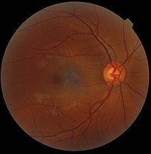
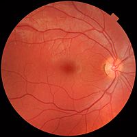
Fundus photographs of the right eye (left image) and left eye (right image), as seen from the front (as if face to face with the viewer).
Each fundus has no sign of disease or pathology. The gaze is into the camera, so in each picture the macula is in the center of the image, and the optic disc is located towards the nose. Both optic discs have some pigmentation at the perimeter of the lateral side, which is considered non-pathological.
The left image (right eye) shows lighter areas close to larger vessels, which has been regarded as a normal finding in younger people.
The fundus of the eye is the interior surface of the eye opposite the lens and includes the retina, optic disc, macula, fovea, and posterior pole.[1] The fundus can be examined by ophthalmoscopy[1] and/or fundus photography.

