Back خيط أكتين Arabic Микрофиламент Bulgarian মাইক্রোফিলামেন্ট Bengali/Bangla གྲུང་སྤྲི་ཉག་ཕྲན། Tibetan Mikronit BS Microfilament Catalan Mikrofilamentum Czech Microffilament Welsh Mikrofilamente German Μικρονημάτιο Greek

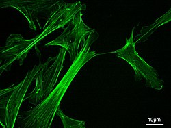
Microfilaments, also called actin filaments, are protein filaments in the cytoplasm of eukaryotic cells that form part of the cytoskeleton. They are primarily composed of polymers of actin, but are modified by and interact with numerous other proteins in the cell. Microfilaments are usually about 7 nm in diameter and made up of two strands of actin. Microfilament functions include cytokinesis, amoeboid movement, cell motility, changes in cell shape, endocytosis and exocytosis, cell contractility, and mechanical stability. Microfilaments are flexible and relatively strong, resisting buckling by multi-piconewton compressive forces and filament fracture by nanonewton tensile forces. In inducing cell motility, one end of the actin filament elongates while the other end contracts, presumably by myosin II molecular motors.[1] Additionally, they function as part of actomyosin-driven contractile molecular motors, wherein the thin filaments serve as tensile platforms for myosin's ATP-dependent pulling action in muscle contraction and pseudopod advancement. Microfilaments have a tough, flexible framework which helps the cell in movement.[2]
Actin was first discovered in rabbit skeletal muscle in the mid 1940s by F.B. Straub.[3] Almost 20 years later, H.E. Huxley demonstrated that actin is essential for muscle constriction. The mechanism in which actin creates long filaments was first described in the mid 1980s. Later studies showed that actin has an important role in cell shape, motility, and cytokinesis.
- ^ Roberts K, Raff M, Alberts B, Walter P, Lewis J, Johnson A (March 2002). Molecular Biology of the Cell (4th ed.). Routledge. p. 1616. ISBN 0-8153-3218-1.
- ^ Gunning PW, Ghoshdastider U, Whitaker S, Popp D, Robinson RC (June 2015). "The evolution of compositionally and functionally distinct actin filaments". Journal of Cell Science. 128 (11): 2009–19. doi:10.1242/jcs.165563. PMID 25788699.
- ^ Kelpsch, Daniel J.; Tootle, Tina L. (December 2018). "Nuclear actin: from discovery to function". Anatomical Record. 301 (12): 1999–2013. doi:10.1002/ar.23959. ISSN 1932-8486. PMC 6289869. PMID 30312531.