Back Neuron Afrikaans Nervenzelle ALS Neurona AN عصبون Arabic خليه عصبيه ARZ স্নায়ুকোষ Assamese Neurona AST Neyron Azerbaijani نورون AZB Neiron BAR
| Neuron | |
|---|---|
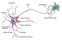
 Anatomy of a multipolar neuron | |
| Identifiers | |
| MeSH | D009474 |
| NeuroLex ID | sao1417703748 |
| TA98 | A14.0.00.002 |
| TH | H2.00.06.1.00002 |
| FMA | 54527 |
| Anatomical terms of neuroanatomy | |
A neuron, neurone,[1] or nerve cell is an excitable cell that fires electric signals called action potentials across a neural network in the nervous system. They are located in the brain and spinal cord and help to receive and conduct impulses. Neurons communicate with other cells via synapses, which are specialized connections that commonly use minute amounts of chemical neurotransmitters to pass the electric signal from the presynaptic neuron to the target cell through the synaptic gap.
Neurons are the main components of nervous tissue in all animals except sponges and placozoans. Plants and fungi do not have nerve cells. Molecular evidence suggests that the ability to generate electric signals first appeared in evolution some 700 to 800 million years ago, during the Tonian period. Predecessors of neurons were the peptidergic secretory cells. They eventually gained new gene modules which enabled cells to create post-synaptic scaffolds and ion channels that generate fast electrical signals. The ability to generate electric signals was a key innovation in the evolution of the nervous system.[2]
Neurons are typically classified into three types based on their function. Sensory neurons respond to stimuli such as touch, sound, or light that affect the cells of the sensory organs, and they send signals to the spinal cord or brain. Motor neurons receive signals from the brain and spinal cord to control everything from muscle contractions[3] to glandular output. Interneurons connect neurons to other neurons within the same region of the brain or spinal cord. When multiple neurons are functionally connected together, they form what is called a neural circuit.
A neuron contains all the structures of other cells such as a nucleus, mitochondria, and Golgi bodies but has additional unique structures such as an axon, and dendrites.[4] The soma is a compact structure, and the axon and dendrites are filaments extruding from the soma. Dendrites typically branch profusely and extend a few hundred micrometers from the soma. The axon leaves the soma at a swelling called the axon hillock and travels for as far as 1 meter in humans or more in other species. It branches but usually maintains a constant diameter. At the farthest tip of the axon's branches are axon terminals, where the neuron can transmit a signal across the synapse to another cell. Neurons may lack dendrites or have no axons. The term neurite is used to describe either a dendrite or an axon, particularly when the cell is undifferentiated.
Most neurons receive signals via the dendrites and soma and send out signals down the axon. At the majority of synapses, signals cross from the axon of one neuron to the dendrite of another. However, synapses can connect an axon to another axon or a dendrite to another dendrite. The signaling process is partly electrical and partly chemical. Neurons are electrically excitable, due to the maintenance of voltage gradients across their membranes. If the voltage changes by a large enough amount over a short interval, the neuron generates an all-or-nothing electrochemical pulse called an action potential. This potential travels rapidly along the axon and activates synaptic connections as it reaches them. Synaptic signals may be excitatory or inhibitory, increasing or reducing the net voltage that reaches the soma.
In most cases, neurons are generated by neural stem cells during brain development and childhood. Neurogenesis largely ceases during adulthood in most areas of the brain.
- ^ UK for neuron; Cambridge Dictionary, https://dictionary.cambridge.org/dictionary/english/neurone
- ^ Najle, Sebastián R.; Grau-Bové, Xavier; Elek, Anamaria; Navarrete, Cristina; Cianferoni, Damiano; Chiva, Cristina; Cañas-Armenteros, Didac; Mallabiabarrena, Arrate; Kamm, Kai; Sabidó, Eduard; Gruber-Vodicka, Harald; Schierwater, Bernd; Serrano, Luis; Sebé-Pedrós, Arnau (Oct 2023). "Stepwise emergence of the neuronal gene expression program in early animal evolution". Cell. 186 (21): 4676–4693.e29. doi:10.1016/j.cell.2023.08.027. PMC 10580291. PMID 37729907.
- ^ Zayia LC, Tadi P. Neuroanatomy, Motor Neuron. [Updated 2022 Jul 25]. In: StatPearls [Internet]. Treasure Island (FL): StatPearls Publishing; 2023 Jan-.
- ^ Cite error: The named reference
Bettswas invoked but never defined (see the help page).